Functions Of Ultrasound During Pregnancy
The images we have of our babies growing in our wombs go beyond mere curiosity to see if they look like mom or if they inherited dad’s nose.
Thanks to the advancement of technology, medicine has found the best impetus to optimize its scope and offer accurate diagnoses and solutions. In the case of pregnancy, obstetricians and perinatologists have in the ultrasounds or ultrasounds great allies to monitor the development of the fetus as if it were just another patient.
I’m sure now you’re wondering what doctors can see through ultrasounds . And to answer you, we will expand a bit, because the functions of this method are diverse.
Ultrasounds are used to rule out various congenital malformations.
According to the week of gestation in which the ultrasound , this will allow to observe in detail the evolution of the fetus and rule out any pathology in time . The idea of this method, which is not invasive or harmful, is to ensure that everything is going well throughout the process.
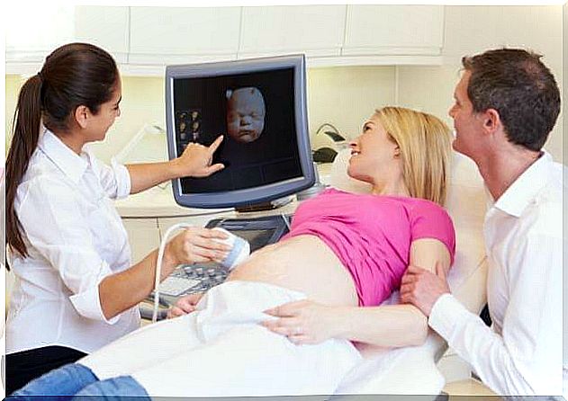
And it is true that mom and dad focus on detailing the baby’s face every time an ultrasound should be performed, but while we stop to appreciate every feature of his face, the doctor takes care of other tests.
What does the ultrasound allow us to see?

The doctor will be in charge of analyzing each aspect of the fetus depending on the week of gestation in which it is. For example , in the first trimester, you will strive to monitor the embryo’s vital signs, such as the heartbeat , and its correct progress to become a fetus.
Starting at week 12, the specialist will look for signs of any congenital disease or malformations . In this phase is that the measurement of the area of the neck of the fetus is practiced, which serves to rule out Down syndrome.
Along with this measurement, the doctor will monitor that the limb developmental milestones and later it will be possible to discover if it is a girl or a boy what anxious parents await.
Through each ultrasound, the doctor will be able to detect any abnormality that could endanger the life of the mother and the baby.
Once the 20th week of gestation is completed, the doctor will be able to see the baby’s internal organs through an ultrasound Doppler , whose image is seen in color and its level of sharpness is greater.
Specialists use the Doppler technique to monitor blood flow through the baby’s arteries and the function of the heart valves , so for many it is very important to have the corresponding equipment in order to obtain a clear picture of everything that happens inside the womb.
Another wonder that can be accessed with an ultrasound is to know the approximate weight and height of the baby.
In the last trimester, the maturity of the placenta and the amount of amniotic fluid will become important.
Among the most advanced procedures, Doppler ultrasound stands out
How many ultrasounds are recommended during pregnancy?
There is no magic number that indicates the number of radiographs that should be performed during the Control prenatal. This by virtue of the fact that it is a non-invasive method, which does not compromise the health of the mother or the fetus, nor does it imply collateral effects.
So the criterion of the treating physician or the protocol of the public or private health system should not be questioned.
In some countries, an ultrasound is done at each prenatal visit, while in other places they prefer to perform only three: one in each trimester of pregnancy.
Talking about 4D ultrasound imaging of the fetus in real time , and with a clarity of volume worthy of astonishment, some doctors recommend its implementation, while others maintain that it is a mechanism that can be dispensed with.
Of course, parents will always take advantage of modernity and want to know in detail the baby that is being formed in the maternal womb , and they will be able to do so if they wish even if the 4D echo is not within the requirements of the doctor who is monitoring the pregnancy.
In short, a correctly controlled pregnancy includes performing the respective ultrasound scans, in which, in addition, parents are offered a printed image that they will be able to keep and thus remember this wonderful stage.